18+ heart diagram easy
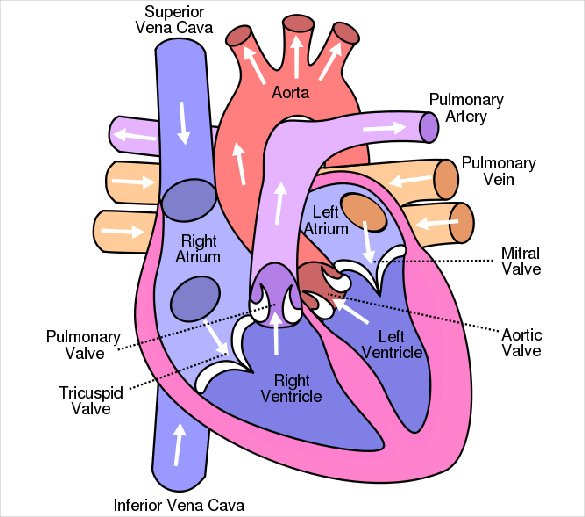
Human Heart Diagram. The right side is in blue and the left side is in red.

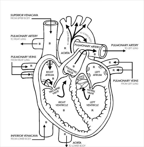
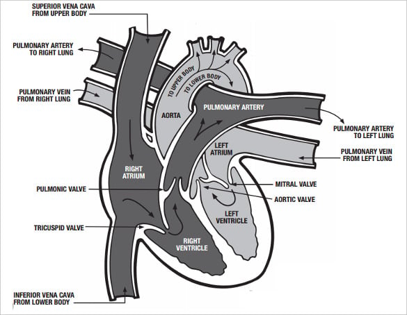
Normal Flow Of Heart Lungs Life After Nclex Rn Nursing Students Cardiac Nursing Nursing School
This is a simple and free human heart anatomy chart for kids.
. The heart though small in size performs highly significant functions that sustains human life. More muscular than the right ventricle. The shape of the human heart is like an upside-down pear weighing between 7-15 ounces and is little larger than the size of the fist.
It is the goal of these nursing mnemonics to provide an easy quick-guide to simplify the concepts of pharmacology. The human heart and its functions are truly fascinating. Featuring six of the basic parts of the heart each numbered and labeled for easy identification.
Blood comes into the right atrium from the body moves into the right ventricle and is pushed into the pulmonary arteries in the lungs. The diagram alongside represents circulation in the human body. The average male heart weighs around 280 to 340 grams 10 to 12 ounces.
The base of the heart is located along the bodys midline with the apex pointing toward the left side. System and the heart. This coordinates with our Human Heart Labeling Worksheet below.
Basic Anatomy And Physiology. Answer the question of why the left ventricle is. A heart diagram is illustrated in several parts so that it is easily understandable to the learners.
The three heart shapes are. Describe the components and functions of the. Pericardium the sac that surrounds your heart.
Myocardium the thick middle layer of muscle that allows your heart chambers to contract and relax to pump blood to your body. This process is called pulmonary circulation. The heart one of the most significant organs.
The heart templates come in varying sizes from large approximately 7 inch size 1 per page medium 5 inch 2 per page small 3 inch 6 per page and mini 2 inch 15 per page. The heart is the organ that helps supply blood and oxygen to all parts of the body. The halves are in turn divided into four chambers.
Blood flows through the heart in 12 easy steps. See more ideas about biology diagrams heart diagram medical drawings. After picking up oxygen the blood travels back to the heart through the pulmonary veins into the left atrium to the left ventricle and out to the bodys tissues through the aorta.
Theres also a page of mixed shapes and sizes from large to mini hearts. Great for USMLE nursing students doctors and medical learners. Human Heart Diagram Drawing class 10.
19 The Cardiovascular System. For better illustration look at the picture below and note how the right and left side are separated. 12 x 18 Ships in 5-7 Days Product ID.
Students usually have to draw diagrams and learn from pictures given in the text book. The heart is made of three layers of tissue. Classic heart tapered heart and rounded heart.
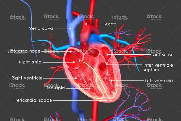
A Labeled Diagram of the Human Heart You Really Need to See. Selecting or hovering over a box will highlight each area in the diagram. Identify the layers of the heart wall.
Explain the events of the cardiac cycle. Because the heart points to the left about 23 of the hearts mass is found on the left side of the body and the other 13 is on the right. This is an online quiz called Nerve Cell Diagram easy There is a printable worksheet available for download here so you can take the quiz with pen and paper.
This amazing muscle produces electrical. The heart is situated at the centre of the chest and points slightly towards the left. Art Print Finished Size.
The heart one of the most significant organs in the human body is nothing but a muscular pump which pumps blood throughout the body. This step-by-step diagram provides easy notes and explanations of the cardiac cycle blood flow through the heart in order and the atrial and ventricular anatomy of the heart. Blood flow through the heart made easy with a simple diagram of the cardiac circulation pathway and steps in order.
Diagram--Which of these vessels returns blood to the left atrium of the heart. Always remember that it must flow through 6 areas on the right side and then 6 areas on the left side this equals 12 steps. Diagram of blood circulation in human body.
On average the heart beats about 100000 times a day ie around 3 billion beats in a lifetime. Made of thin layers of tissue it holds the heart in place and. Conducting system of the heart.
When viewing a dissected heart it is easy to visually discern the right and left ventricles by _____. Endocardium the thin inner lining of the heart chambers that also forms the surface of the valves. Posted on June 8 2016 by admin.
So the partial pressure of. How does blood flow through the heart step by step quizlet. This free crochet heart pattern is super easy and fun to make and works up extremely quickly.
If you want to redo an answer click on the box and the answer will go back to the top so you can move it to another box. The inferior tip of the heart known as the apex rests just superior to the diaphragm. Sep 21 2019 - Explore Tehreem Liaqats board Diagrams on Pinterest.
It is divided by a partition or septum into two halves. The heart pumps around 57 litres of blood in a day throughout the body. This quiz has tags.
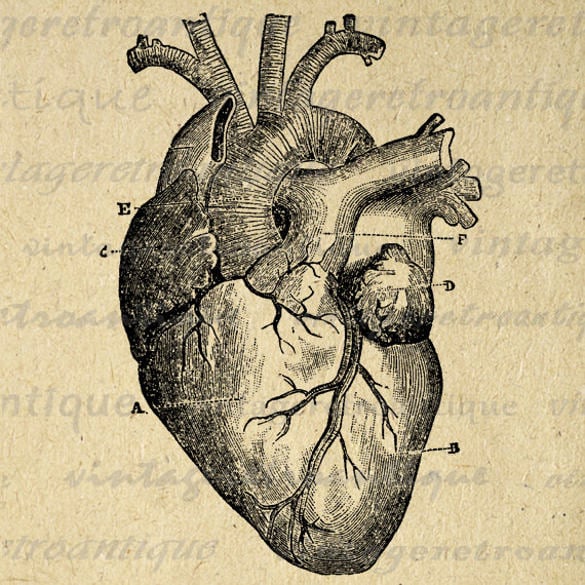
Human Heart Labeling Chart. Drag and drop the text labels onto the boxes next to the heart diagram. 53762063003A Related Tags Categories Vintage.
Click on the tags below to find other quizzes on the same subject. The heart is situated within the chest cavity and surrounded by a fluid-filled sac called the pericardium. In this interactive you can label parts of the human heart.
The shape of the human heart is like an upside-down pear weighing between 7-15 ounces and is little larger than the size of the fist. Describe the general features of the heart. Heart anatomy video quiz and chart included.
You can use different yarn weights to create hearts of varying size but just to give you an idea of the size all the hearts shown in this post are crocheted with cotton thread size 10 and a 2 mm crochet hook and they measure about 45 by 45 cm 177 by 177 inches. The cardiac cycle has 2 phases systole and diastole defined by depolarization and contraction vs repolarization and relaxation. AP II Chapter 18 Heart.
This diagrammatic representation of human body parts makes it easy for science students to learn about the functionality and working of the organs. It is located between the lungs in the middle of the chest behind and slightly to the left of the breast bone. Four Chambers of the Heart and Blood Circulation.
13 Heart Diagram Templates Sample Example Format Download Free Premium Templates
Heart Diagram 15 Free Printable Word Excel Eps Psd Template Download Free Premium Templates

Http Www Theharleystreetcardiologypractice Com Wp Content Uploads 2014 04 Eps Gif Medical Knowledge Ekg Interpretation Heart Pictures

13 Heart Diagram Templates Sample Example Format Download Free Premium Templates

Pin Em Heart

Heart Diagram Diagram Picture Heart Diagram Cardiac Nursing Nursing Study
![]()
13 Heart Diagram Templates Sample Example Format Download Free Premium Templates
13 Heart Diagram Templates Sample Example Format Download Free Premium Templates

Basic Heart Diagram Worksheet
13 Heart Diagram Templates Sample Example Format Download Free Premium Templates

Pin On A Pi

Easily Remember The Most Common Cause Of Right Sided Heart Failure Nursing School Survival Nursing School Studying Nursing Students

Electrical Conduction System Of The Heart Normal Intrinsic Electrical Conduction Of The Heart Allows Electr Fast Heart Rate Atrioventricular Node Heart Rate

Anatomy Of The Human Heart Cardiology Educational Poster 24x36 Medical Anatomy Cardiology Human Heart
Heart Diagram 15 Free Printable Word Excel Eps Psd Template Download Free Premium Templates